
In How Is a Varicocele Treated?, we discussed the shared principle behind five common approaches: rather than trying to repair a poorly draining vein, the procedure closes it so blood can use other return pathways.
This article focuses on one high route, formally named a “minimally invasive high ligation procedure for interrupting reflux”; below, we use “high ligation” to explain the design logic for addressing the reflux source. For a side-by-side overview of operative locations and general considerations, return to the neutral comparison of five options.
Why work at a “high” level?
The left testicular vein travels upward and joins the left renal vein. The high-route approach addresses the reflux source near this junction along the main return pathway, allowing blood to use other routes, such as the deep pelvic veins and the femoral vein, before returning to the larger venous system.
Compared with a lower location, the venous branches have converged more at this level. Our existing treatment-options guide describes about 4–6 vessels at the high level, compared with about 8–12 venous branches that may be identified in the low microsurgical route. This is an anatomical difference between operative levels; it does not mean every person has the same number of vessels or the same operative plan.
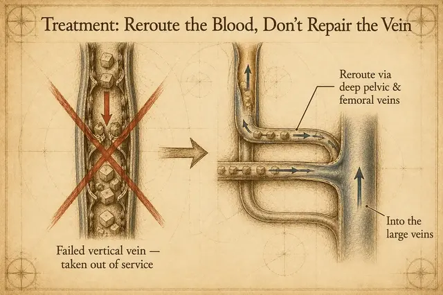
How can a varicocele appear again? Unaddressed branches matter
Reflux in a varicocele is not limited to one main trunk. If a branch remains unaddressed, reflux pressure may persist. Blood can still back up into the pampiniform plexus, and the dilated appearance or discomfort may appear again.
From a design perspective, working where there are relatively fewer branches aims to concentrate the routes that need to be identified and addressed, reducing the opportunity to miss a branch. This describes the mechanism, not an individual outcome. Vessel anatomy, decisions made during the procedure, and follow-up all remain relevant for each person.
With bilateral findings, why is the left side often assessed first?
The right testicular vein has a shorter return path and drains directly into the inferior vena cava. The left testicular vein joins the left renal vein and is more likely to face higher return pressure. There are also communicating branches between the two testicular venous systems: when reflux pressure on the left rises, the right side may also become congested, producing a bilateral finding.
When assessment indicates that the main reflux source is on the left, the physician may first consider taking the left reflux route out of service and assess whether the right side changes as the pressure changes. If an individual's anatomy and findings call for it, both sides may be planned in the same procedure. This does not mean that every bilateral case is managed on the left alone; ultrasound findings, symptoms, and operative observations all matter.
What the procedure looks like: questions worth clarifying
This route uses an incision of about 2.5–3 cm. The precise incision, anesthesia plan, going-home arrangements, and pace of returning to activity differ with a person's health and operative plan. In general, ask the operating physician before surgery about the expected scope, activity precautions, and follow-up; after surgery, follow that physician's care instructions.
Risks, limitations, and suitability
As with any operation, this procedure carries risks of bleeding, infection, and anesthesia-related complications; how these apply to you depends on your individual health, and they are worth discussing with your physician before surgery.
The high route also has operative limits: distinguishing artery from vein at a high level is not easy, and care is needed to avoid ligating the testicular artery — if the artery is affected, the blood supply to the testicle may be compromised. If lymphatic vessels are ligated along the way, fluid can collect around the testicle after surgery (a hydrocele). The surgical field, working space, and anesthesia plan also need consideration. Even after venous treatment, a varicocele can recur — for example, venous collaterals that branch off below the interception point cannot be addressed from the high level and may allow reflux pressure to persist. High ligation is not automatically suitable whenever dilated veins are present: the physician needs to assess the reflux location, vessel anatomy, symptoms, test findings, and overall health.
When the source of symptoms is not yet clear, other scrotal or inguinal conditions still need to be distinguished, or individual clinical factors and anatomy do not fit this route, the physician may discuss further evaluation or another management approach. Understanding these limits before surgery is as important as understanding the mechanism.
Key takeaways
- High ligation addresses the main reflux route at a high level near the left testicular vein's junction
- About 4–6 vessels are described at the high level, while about 8–12 venous branches may be identified in the low microsurgical route; this reflects anatomy at different levels
- An unaddressed branch may let reflux pressure persist and is one mechanism by which a varicocele can appear again
- Bilateral findings do not necessarily mean that both sides have independent reflux sources; left-sided pressure and communicating branches need to be considered
- Suitability, whether both sides need attention, and the recovery plan should be discussed for the individual case
A final word
If you are considering treatment for a varicocele, bring ultrasound or other test reports, notes about when symptoms occur, and your questions about anesthesia and daily arrangements to the appointment. Asking where the reflux is, which branches may need attention, and why the plan is arranged this way can help you and your physician make a decision that fits your situation.