
In What Is a Varicocele?, we discussed why the pampiniform plexus becomes enlarged and a varicocele develops. It is mainly related to three factors:
- The testicular vein facing backflow pressure at the high junction point
- The "nutcracker effect" of the left renal vein being pinched by the aorta and the superior mesenteric artery
- Incompetent valves within the spermatic veins
So how is a varicocele treated? This article starts with the underlying principle, then outlines several procedures commonly used in clinical practice, so you have a basic understanding of the options before your appointment.
The treatment principle: "take the longer route" and reroute the blood
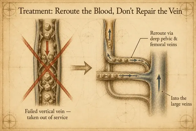
Returning to the analogy we used before — think of a vein as a conveyor belt and venous blood as the cargo. The original "left testicular vein" is a vertical, upward-running belt. It's the shortest path, but because it has to work against gravity, once the gates (valves) fail, the cargo backs up and accumulates.
The idea behind treating a varicocele is counterintuitive: rather than repairing this failed belt, we simply take it out of service.
Once it's out of service, the cargo (venous blood) automatically reroutes onto nearby horizontal belts — such as the deep pelvic veins and the femoral vein in the thigh. The path is longer, but because it runs horizontally and doesn't have to fight gravity, it actually flows more smoothly. These horizontal belts ultimately merge into the large venous system, drawn back to the heart by the suction of the main lines.
Once the vertical, hard-to-travel, broken belt is decommissioned, blood no longer backs up where it was, the dilated veins gradually regress, and symptoms improve along with them.
So nearly every "varicocele surgery" does the same thing: it closes off the failed vein. What differs is "where it's closed," "how it's closed," and "from which approach you enter." That's what gives rise to the several procedures commonly seen in clinical practice today.
Five common procedures
The following are organized by "how low or high the vein is closed off" and "what method is used to close it." Each differs in its principle, location, and general considerations; how to choose in practice is discussed with your physician based on your individual situation.
1. Microsurgical subinguinal varicocelectomy
An incision of about 2–3 cm is made at the "low inguinal" position. Under high-magnification microscopy, the spermatic cord and the surrounding veins are located, and the venous branches identified during surgery are ligated one by one (usually about 8–12 of them).
Principle: Closing off the branches leading to the higher veins from a low position near the source, so the blood reroutes.
Operative considerations:
- Working under the microscope, the surgeon must distinguish artery, vein, vas deferens, and lymphatics
- Care is taken to avoid ligating the artery (to avoid affecting testicular blood flow) and the lymphatics (to avoid post-operative scrotal edema)
- Surgery takes around 2 hours and relies more heavily on microsurgical training
- Different academic studies discuss recurrence rates, post-operative semen parameters, and similar measures quite extensively; actual outcomes relate to the surgeon's experience
2. Inguinal varicocelectomy (Ivanissevich approach)
Positioned slightly higher than the microsurgical procedure, an incision of about 3–5 cm is made at the "inguinal" level. The veins within the spermatic cord are located — magnifying loupes may help — and the thicker spermatic veins are ligated one at a time (usually about 3–8 of them).
Principle: Closing off after the branches have merged into several medium-sized veins, with more working space than the microsurgical technique.
Operative considerations:
- At this level the veins are already thicker than at the low position, making visual identification relatively easier
- Some deeper or lateral small branches may not join the spermatic cord, and the limited field of view means they can't always be fully intercepted
- Surgery takes around 1–2 hours
3. Suprainguinal / high ligation (Palomo approach, retroperitoneal route)
Closing off at a higher position — ligating the main trunk directly, just before the testicular vein joins the left renal vein.
Principle: The main return pathway can be closed off at a higher position. The number of vessels needing ligation is usually only about 4–6 (on the left).
Historical background: Procedures based on this principle came into wide use from the mid-to-late 20th century (such as the retroperitoneal route proposed by Palomo) and were long one of the main options.
Operative considerations:
- The incision is larger and involves cutting through the abdominal wall muscle, which may affect muscle recovery or produce other complications
- Distinguishing artery from vein at this high position is not easy, and care is needed to avoid ligating the testicular artery
- The surgical field and space are limited, so general or spinal anesthesia is usually required
4. Laparoscopic varicocelectomy (high ligation)
A laparoscope (endoscope) is used to view the testicular vein from inside the abdominal cavity, which is then managed with vascular clips or energy instruments.
Principle: The same closure location as "high inguinal ligation," but using a laparoscopic view and instruments instead.
Operative considerations:
- The course of the vessels can be observed through the laparoscope
- The abdominal-wall wounds are smaller (usually 3–4 incisions of about 0.5 cm)
- The abdomen must be insufflated to expand the cavity, so it's generally performed under general anesthesia
- Because surgery is performed from inside the abdomen, it involves contact with the intestines and other intra-abdominal organs; over the long term there's a small possibility of bowel adhesions
5. Percutaneous embolization
No open surgery is required; this is performed by an interventional radiologist. A small incision is made in the right groin and a catheter is introduced, passing through the femoral vein, the inferior vena cava, and the left renal vein, finally reaching the left testicular vein, where a sclerosing agent or embolic material is injected to block off the vein.
Principle: Blocking the high vein from inside the vessel, achieving the same closure location as "high ligation" but without a surgical incision.
Operative considerations:
- Usually performed under local anesthesia, with a typically shorter hospital stay
- Developed since the 1980s, it has long been one of the options used by urology and interventional radiology
- The procedure uses X-ray fluoroscopy, so both physician and patient are exposed to radiation
- There is a possibility of the sclerosing agent / embolic material leaking to other locations
- Its suitability for a given case is discussed jointly by interventional radiology and urology
How to choose? There's no single "best" answer
By this point you'll have noticed that all five methods actually do the same thing — closing off the failed vein, at different locations and with different tools.
In practice, the choice is made by the doctor weighing the following factors together:
- The varicocele's grade (Grade 1/2/3) and the severity of symptoms
- Whether there are fertility considerations, and the results of any semen analysis
- The patient's age, lifestyle and type of work, and anesthesia considerations
- Whether there are accompanying conditions (hernia, hydrocele, a prior history of inguinal surgery, and so on)
- The doctor's own training background and the techniques they're experienced with
Each technique has its own strengths and limitations; the academic literature has no absolute consensus on a "best technique." Most comparative studies discuss them using different measures — recurrence rate, post-operative pain, complications, post-operative semen parameters — and the conclusions of different studies don't entirely agree.
Key takeaways
- The thinking behind treatment isn't to "repair" the vein but to "decommission" the failed one and let the blood reroute
- The five common techniques seen in practice all do the same thing; what differs is the closure location and the method
- Microsurgery, inguinal ligation, high ligation, laparoscopic ligation, and embolization each have their own characteristics and suitable situations
- There's no single "best" method — the choice is made by weighing your individual situation, the doctor's recommendation, and patient preference together
A final word
Faced with so many options, the most important thing isn't to pick the "best" one yourself, but to bring your test reports, a record of your symptoms, and the things you care about (for example: length of recovery, fertility plans, anesthesia considerations) to a urology or general surgery clinic and discuss them fully with your doctor.
Every technique has its own design logic and the kind of patient it suits. Letting a professional assess your individual situation is more reassuring than deciding from a single article alone.